
Localization Studies for Hyperparathyroidism
Hyperparathyroidism is an unusual disease, caused one or more of the parathyroid glands becoming over-active and producing too much hormone (PTH). Sometimes the gland that is overworking becomes large and produces a lot of PTH. Other times the gland grows only minimally and produces just a little more PTH then expected, but the person’s body maybe sensitive and this little extra PTH can create chaos in the body. So the diagnosis of hyperparathyroidism is almost always made by looking at the relationship between calcium, PTH & vitamin D. The scans are not used to make the diagnosis of hyperparathyroidism, your doctor depends on lab tests to confirm that you have hyperparathyroidism.
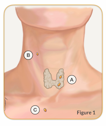
This is different then most other diseases in the body. To add to this complexity, develop in the throat of the baby that is in the mother’s belly, and travel down into the neck. This movement of parathyroid can be problematic because only 80% times the parathyroid come to finally be positioned behind the thryoid (position A in Figure 1). The other 20% of times it can travel very little and be in the throat, or travel too much and end up in the chest (position B & C in Figure 1). 3% of people who only 3 parathyroid glands, and 10% have more then 4. So once the diagnosis of hyperparathyroidism is made Dr. Larian will use a parathyroid scan to help identify which parathyroid gland is abnormal, and where it is located.
This is all done before the day of your surgery so that the doctor to plan your surgery in advance and clearly discuss it with you. There are many parathyroid scan options, and choosing the right one is sometimes difficult. Dr. Babak Larian, prefers ultrasound of the neck as the first scan to localize the abnormal parathyroid gland. This is because it has a high chance of finding the abnormal gland without exposing the person to any type of radiation.
Ultrasound
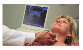
Ultrasound imaging is a reliable way to identify an abnormal parathyroid gland. It uses harmless soundwaves to look at the structures under the skin, including the thyroid, parathyroid, muscles, and blood vessels. An ultrasound should always be the f irst study to look for an abnormal parathyroid because it is very accurate but does not expose a patient to any type of radiation. When an ultrasound is performed by an expert radiologist, endocrinologist, or surgeon, it has a high likelihood to show an abnormal parathyroid.
In the past decade, ultrasound has become available in many doctor’s offices around
the world, and most expert parathyroid surgeons have an ultrasound machine and can
immediately do a scan for a patient when HPT diagnosis is confirmed. In this way the
patient can start treatment right away.
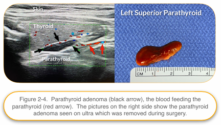
A parathyroid ultrasound can show the exact dimensions of the thyroid gland, enlarged parathyroid glands with the vessels feeding it, carotid artery, lymph nodes, the portion of the thymus that is in the neck, breathing tube (trachea), and the surrounding muscles. The parathyroid adenomas on the 3 ultrasound images show parathyroid adenomas of varying shapes and sizes; and as you can see when the parathyroid is removed in surgery the shape and size directly correlates to f inding on ultrasound.
Additionally, the surgeon can see exactly where the abnormal enlarged parathyroid is with respect to the thyroid gland, deep to it (superior parathyroid) or in the case of the inferior parathyroid just below the lower end of the thyroid gland.
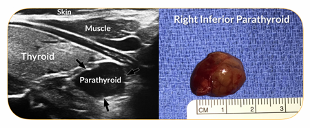
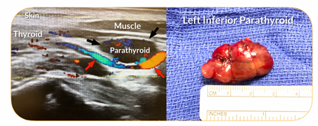
You can also tell how deep the abnormal gland is from the skin, and how big are the blood vessels that are feeding the parathyroid.
Another important benefit of ultrasound is that it is by far the best scan to show the thyroid gland itself. The surgeon can see if there are any nodules in the thyroid gland that need to be addressed at the same time as parathyroid surgery. In the rare case of a parathyroid hiding inside the thyroid gland, the ultrasound would show it. An ultrasound does have its limitations; it cannot be used to show what’s behind bone or cartilage. So, if a parathyroid adenoma is hiding behind the collarbone, chest bone, Figure 5 breathing tube, or voice box, other parathyroid scan options may need to be considered.All expert parathyroid surgeons perform their own ultrasound to confirm the size and location of an abnormal parathyroid and assess the relationship of the abnormal gland to adjacent structures (thyroid, carotid artery, and recurrent laryngeal nerve), as well as the distance away from the skin.

This information is helpful in planning for parathyroid surgery. In cases when both an ultrasound and sestamibi scan have been done to identify an abnormal gland in the same area, the accuracy of the findings is greater than 97%. Dr. Larian repeats the ultrasound in the office to determine the precise location of a parathyroid adenoma so that its exact location is identified to minimize risk during surgery further.
PROS – 1. Very accurate. 2. Can show the anatomy of the neck very well and create a roadmap for a surgeon to use to access one or more abnormal parathyroids and avoid important structures. 3. There is no radiation exposure for a patient. 4. Can be done in an office
CONS – 1. Hard for a surgeon to visualize the anatomy when the procedure is done at another imaging center and not by the surgeon. 2. Does not show parathyroids that are hidden behind the voice box, breathing tube, chest bone, or collarbone. 3. Patients that have thyroid inflammation (Hashimoto’s thyroiditis) will often have lots of enlarged lymph nodes that can be confused on a parathyroid ultrasound for an abnormal parathyroid gland
Parathyroid Scan FAQ
Dr. Larian understands that patients often have a lot of questions about the imaging tests required for locating parathyroid tumors and that the choice of which scan to get can be confusing. He has answered some common questions for you below to help you through this process.
Can localization scans help make the diagnosis of hyperparathyroidism?
No, the diagnosis of HPT is made by laboratory findings. The scan is only to help identify where the abnormal gland is located.
Which scan should be the first choice?
Ultrasound should always be the first choice because it only uses soundwaves. There is no radiation involved during an ultrasound, too.
Should I be concerned about radiation exposure?
Many patients express some concern in regards to being exposed to radiation for parathyroid scans. First, the decision to proceed with one of the studies that uses radiation (sestamibi, SPECT, or 4D CT scan) should only be made if the ultrasound is not successful in finding the abnormal parathyroid. Having said that, the dose of radiation in sestamibi, SPECT, and 4D CT scans is low and generally not harmful. Knowing the exact location of the abnormal parathyroid allows a surgeon to perform a minimally invasive focused surgery, which limits scarring, trauma to tissue under the skin, and recovery time. This outweighs the disadvantage of radiation exposure of a scan.
Can a single parathyroid scan be used to identify an abnormal parathyroid gland?
At the CENTER, Dr. Larian is able to find a great majority of abnormal parathyroids on ultrasound alone. A parathyroid surgeon performing an ultrasound has the added advantage of being to able to compare ultrasound findings to what they see in surgery; this in time gives the parathyroid surgeon an exceptional amount of insight. A minority of patients require multiple localization studies to identify an abnormal parathyroid gland. Each localization study is used to help create a personalized treatment plan for a patient, and ultimately, ensure that a patient can alleviate HPT symptoms.
If a parathyroid scan shows that a patient is dealing with an abnormal parathyroid gland, what happens next?
Once an abnormal parathyroid is identified, then a focused surgery to remove the abnormal parathyroid can be done.
What will happen if a parathyroid scan comes back negative?
Negative results from a localization study only means the abnormal gland was not visible on that particular scan, NOT that the person does not have hyperparathyroidism. Additional tests need to be find the abnormal parathyroid gland.
When do the results of a parathyroid gland scan become available?
Ultrasound done in the office will give you results immediately. Otherwise scans done at imaging centers/hospitals will give results in 1-2 days. Fortunately, at CENTER, Dr. Larian is able to get results of scans from the imaging center or hospital we use on the same day, and in fact can bring up the images and review them with the patient right away.
How long does it take to complete a parathyroid gland scan?
The length of time required to complete a parathyroid gland scan varies from a few minutes for ultrasound & CT scan, to 45 minutes for MRI, and hours for Sestamibi SPECT scan.
Contact The CENTER For Advanced Parathyroid Surgery Today
Dr. Larian is an experienced parathyroid surgeon in Los Angeles with over 2 decades of experience in hyperparathyroidism surgery. unique knowledge in parathyroid gland diagnostic testing and the meaning and ramifications of parathyroid gland testing for patients suffering from HPT. Dr. Larian is happy to perform localization studies to help a patient diagnose HPT. And if a patient is dealing with HPT, Dr. Larian can provide a custom treatment recommendation. If you feel as though you may be experiencing parathyroid problems and would like to speak with an expert parathyroid surgeon today, we encourage you to contact our CENTER by calling 310.461.0300 to set up an initial consultation.